Ants are a lot of things. They’re nearly everywhere you look. They’re abundant — at least 20 quadrillion at last estimate. Some wreak havoc. Some are just weird. And many are crucial to the health of their environments.
“They have interesting social structures and complex societies,” added Evan Economo, a biodiversity scientist at the University of Maryland.
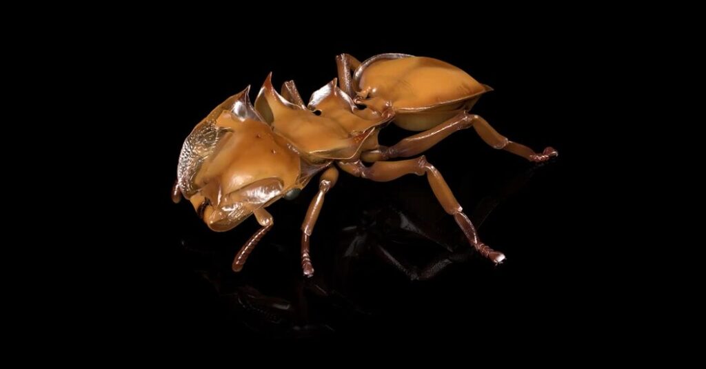
And now, in a study published on Thursday in the journal Nature Methods, ants are the stars of a stunning parade of high-resolution 3-D images made by Dr. Economo and his colleagues. The scans reveal the insects’ diverse bodies, inside and out, in exquisite anatomical detail. The entomological catwalk is also available for free at antscan.info.
Nearly 2,200 specimens, drawn from museums and personal collections around the world, were X-ray scanned in a single intense week with a synchrotron particle accelerator in southwest Germany. Traditional, smaller-scale methods of making such scans would have typically taken years.
Several people worked two shifts around the clock, including Thomas van de Kamp, a biologist based at the Karlsruhe Institute of Technology that housed the accelerator. “The diversity is absolutely stunning. There’s very huge ones like the bullet ants with their most painful sting in the insect world,” he said. And “we scanned super tiny ants — specialist predators of spider eggs, for example.”
Smaller micro-CT scanners, including the kind used for years in Dr. Economo’s lab, work well to create 3-D images. But they’re slow. A synchrotron can scan samples more quickly because it’s exposing them to far more light. And it offers the ability “to visualize the soft tissues without the need for” adding a contrast agent for staining the samples, Dr. van de Kamp said.
The initial renderings replicated the contorted postures of many of the preserved ants. “They’re kind of all crinkled up,” Dr. Economo said.
So his computer science collaborators used artificial intelligence to automate the repositioning of the scans to give them more natural poses. He believes the technique, which he hopes might one day include all living things on Earth, can be used by scientists, educators and even artists and animators.
Dr. Economo began the collaboration with Dr. van de Kamp when he was at the Okinawa Institute of Science and Technology in Japan. Julian Katzke, then a graduate student in Dr. Economo’s lab, recalled specimens pouring in from all over the world. Dr. Katzke then flew to Germany with more than 2,000 vials packed inside two tote bags.
“Even though it’s many specimens, they all still fit in a suitcase,” he said. “So that’s a benefit of working on small insects.”
Inside the synchrotron, a robot positioned sample after sample for scanning. Dr. van de Kamp looked on with fascination from the control room, watching as these insects materialized on the screen in front of him. “For me, it was just amazing how much they differ,” he said, considering all the “many peculiar-looking ant species with specialized mandibles, spines or hairs.”
The scans also allowed the researchers to peer inside the tiny ant bodies, revealing their brains, guts and glands.
Once the team had the 3-D images in hand, they demonstrated their power. For instance, researchers previously knew that the exoskeleton of one type of fungus-farming ant is a kind of mineralized armor, “almost like the shell of a mollusk,” Dr. Economo said. The new scans allowed a rapid survey of many additional species, and they found the armor was common among a variety of other ants from the same group.
Jessica Ware, curator and chair of invertebrate zoology at the American Museum of Natural History who wasn’t involved in the study, found the new work transformative. “I don’t use this word lightly,” she said.
Dr. Ware believes researchers will now be able to look inside their valuable study organisms without ever having to slice them open. And despite the costs of using a synchrotron, she’s confident that the approach will be adopted more widely.
Dr. Economo is also excited about broadening the approach beyond ants “where we can do everything from build digital worlds populated with realistic organisms to design robots,” he said.
And he remains committed to the idea of allowing the public access to these detailed models of organisms that, in his words, “reflect the complexity of shapes that you see in nature.”
The post You’ve Never Seen Ants Like This Before appeared first on New York Times.