The patients were old — more than 2,200 years old. But the medical experts were determined to give them a cutting-edge 21st-century exam.
First up was Nes-Hor, a priest in the Temple of Min, who died circa 190 B.C.E. and whose body was wrapped in a linen shroud that had blackened over the centuries. Then came Nes-Min, circa 330 B.C.E., who had been draped in a netted garment with strands of vibrant beads.
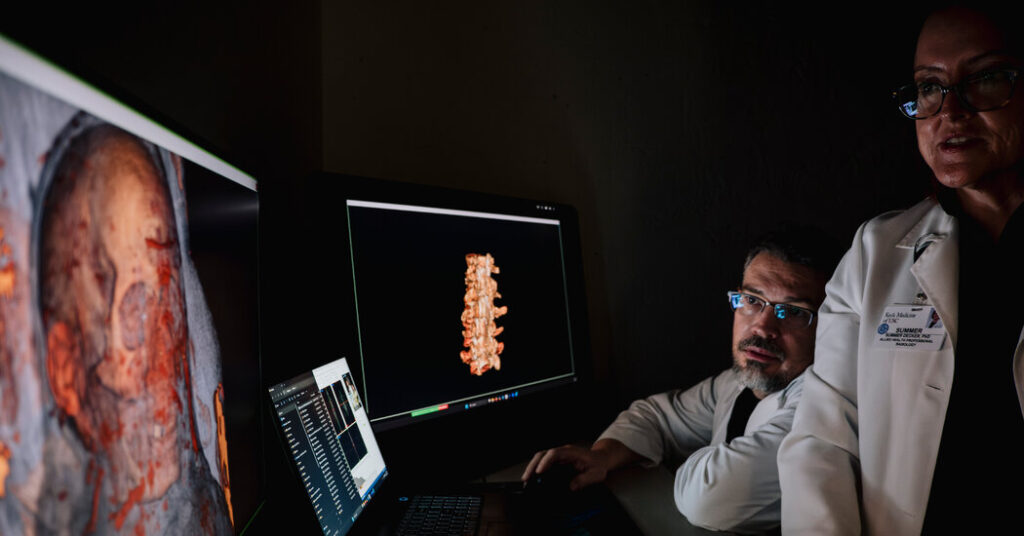
Researchers at the University of Southern California recently used high-resolution CT scanners and medical-grade 3-D printers to conduct virtual autopsies on the two Egyptian mummies. Their goal, as with any patient, was to illuminate ailments and injuries.
The scanner captured 320 different cross-section images of the mummies per rotation, slices that stacked together “like a loaf of bread” to form 3-D digital models, said Summer Decker, the director of the university’s Center for Innovation in Medical Visualization, who oversaw the project. From there, her team analyzed the mummies’ various anatomical structures and used 3-D printers to create life-size reproductions of their spines, skulls and hips.
Radiology is a fast-moving field, and “as technology advances, you’ve got to go back and look, and ask what you might learn from your new tools,” Dr. Decker said. Given the high resolution — the slices were less than half a millimeter thick — the team was able to find artifacts and details that were new or even contradictory to past reports.
Researchers had previously noticed, for example, that Nes-Min, who they believe lived into his 40s, had broken bones along his right rib cage, all of which had healed, suggesting some type of traumatic fall or attack he had survived earlier in life. They also believed he suffered from chronic lower back pain, given that he had a collapsed lumbar vertebrae. Dr. Decker and James Schanandore, a human anatomist who studies prehistorical remains, discovered possible burr holes in the spine, which suggested to them that he had most likely undergone some type of back surgery similar to trephination, which was almost unheard-of at the time.
“It’s interesting to see some of the same diseases that our modern populations have,” Dr. Decker said.
Past reports had also indicated that Nes-Min probably died of a dental abscess, but the new high-resolution models did not show evidence of something serious enough to be fatal.
The scans of Nes-Hor, who lived to be about 60, revealed the intricate details of a severely deteriorated hip, which researchers believe would have caused a serious limp.
“When people can get beneath the surface of these mummies — let them see the source of the back pain or the hip pain — people can see them not as exotic artifacts but as human beings,” said Diane Perlov, an anthropologist and the head of exhibits with the California Science Center, where the mummies and their prints will be on display starting Feb. 7. “It’s really an emotional experience.”
The medical-grade 3-D printers are the same technology that surgeons use to transform M.R.I. and CT scans into physical models that they can practice on, better envisioning the size of a patient’s tumor or a malfunction inside a patient’s cardiac passages. Doctors also sometimes use the prints to help patients better understand their own conditions and treatment plans, letting them hold an exact replica of their own organ in their hands.
In the case of the mummies, Dr. Decker and her colleague, Jonathan Ford, also printed replicas of artifacts that were inside the sarcophagus, including ceremonial scarab beetles and clips that might have held the mummy wrapping in place, much like the metal clasp used to secure an ACE bandage today. Those artifact replicas, which can be printed in five million color options, allow scientists to handle them without unwrapping the mummies and risking any damage.
But most astounding to Dr. Perlov was the lifelike detail in the soft tissues and facial features, including the eyeballs, eyelids, ears and lips. “It’s incredible,” she said, reviewing the scans.
“What we’re trying to do,” Dr. Decker said, “is go beneath the layers of all that wrapping, and see that this was a living person who had their own problems.”
Emily Baumgaertner Nunn is a national health reporter for The Times, focusing on public health issues that primarily affect vulnerable communities.
The post What Do You Get When You Put a Mummy Through a CT Scan? appeared first on New York Times.